
High-frequency Ultrasound Imaging for Cardiac Fibers
3D Ultrasound and MR Diffusion Tensor Imaging
Simulated Ultrasound Image from MR Diffusion Tensor Image
Cardiac Fibers in Pig Hearts
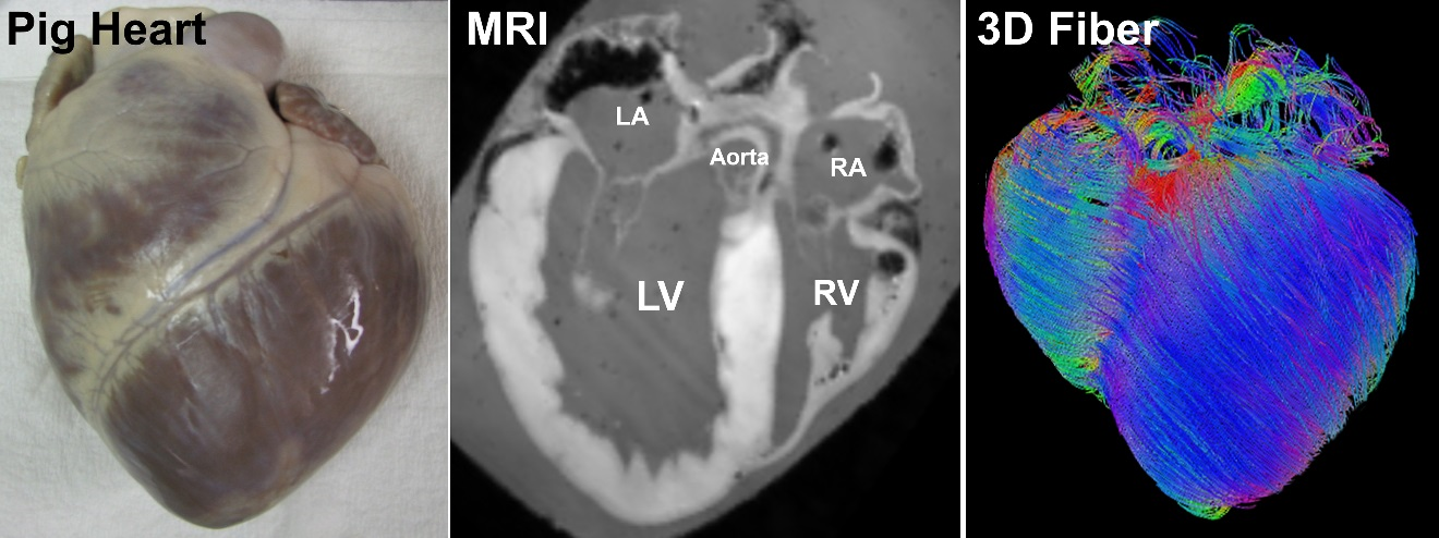
Cardiac fiber imaging of a pig heart. By adding new clinically valuable information on cardiac fibers, heart modeling, and strain measures to widely-used echocardiography, our research is to deliver an innovative, noninvasive imaging tool for cardiac examinations, which will have significant impacts on the healthcare and clinical management of millions of patients with heart failure.
We demonstrated our fiber mapping method in pig hearts where the size of the pig heart is close to that of a human heart. We have applied our fiber mapping tool to the hearts of mice, rats, canines, and pigs. We used animals to study cardiac fibers in both in vivo beating hearts and ex vivo excised hearts for rigorous validation, which is difficult in human patients. When translating the method to human, we consider the following aspects. i) 3D Ultrasound. The principle of estimating fiber orientation has been tested on human hearts using MRI/CT. Our method uses 3D ultrasound instead of CT or MRI because ultrasound has wide applications in cardiac examinations and because 3D ultrasound is commercially available for routinely clinic examinations. ii) Heart size and ultrasound scanner. Our fiber mapping tool can be applied to a heart of any size if its 3D ultrasound images and fiber atlas models are available. For our pig heart experiment, we used our 3D scanning setup and a clinical ultrasound system, which is routinely used in human cardiac studies. Therefore, the heart size and ultrasound scanner are applicable to human patients. iii) Fiber atlas models. Our fiber mapping tool is able to take different atlas models as the input. The NIH-supported Cardiac Atlas Project, an international collaboration, established a large-scale standardized database of cardiac imaging examinations and derived functional analyses. High-resolution atlas data of human hearts are publically available at multiple sources.