High-field Magnetic Resonance Imaging (microMRI)
High-resolution Small Animal PET/CT (microPET/CT)
Multispectral In Vivo Fluorescence Imaging
A limiting factor for in vivo fluorescence imaging is the confounding signal from the autofluorescence of the animal. Our multispectral imaging system utilizes full multispectral imaging and spectral unmixing algorithms; thus it is able to separate the tissue autofluorescence from the signal of interest, thereby greatly increasing sensitivity and signal-to-noise. The multispectral in vivo fluorescence imaging system could be used to guide the development and evaluation of new drugs, nanoparticles, quantum dots, and imaging probes. Therefore, this device would provide synergy among clinical research involving multiple emphasis areas: imaging, nanotechnology, cancer, the drug discovery, inflammation/vaccines, and animal models of human disease.
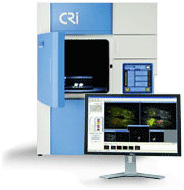
This system is located at the Quantitative BioImaging Laboratory (QBIL) in Wesley Woods Health Center, 2nd floor.
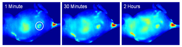
Fluorescence images of a tumor-bearing mouse after being injected with gold nanoparticle-conjugated photodynamic therapy drug at a) 1 min, b) 30 min, c) 120 min after intravenous tail injection. Unprecedented delivery efficiency and accumulation rate of the drug in the tumor is monitored via its fluorescence increase in the tumor area (white circle). Cheng Y, C Samia A, Meyers JD, Panagopoulos I, Fei B (co-corresponding author), Burda C. "Highly efficient drug delivery with gold nanoparticle vectors for in vivo photodynamic therapy of cancer". J Am Chem Soc. 2008 Aug 13;130(32):10643-7 Abstract|Full Text