Image Registration
Image Classification
Image Segmentation
Attenuation Correction
Partial Volume Correction
Motion Correction
Positron emission tomography (PET) is able to differentiate between benign and malignant tumors, to determine tumor stages, and to assess tissue response to therapy. However, despite continuing improvement of the physical instrumentation of PET, it is stilled limited by low spatial resolution compared with that of high resolution magnetic resonance imaging (MR) or computed tomography (CT). The imaging physics, the detector design, and the reconstruction procedure impose a theoretical limitation on PET resolution. The low spatial resolution results in a PET partial volume effect which consists of two distinct phenomena. The first is the tissue fraction effect resulting from the relatively low spatial resolution of PET where a voxel could consist of different structures and could undermine the subresolution tissue heterogeneity. The second is the point spread function (PSF) blurring of PET imaging. The response of a PET scanner to a point source activity shows a bell shape instead of a perfect impulse function. This point spreading effect causes activity spillover between image pixels so that the imaged activity at a voxel is not only from the source at the location but also from activity in its neighboring regions. The PET partial volume effect largely biases the quantitative measurement of the regional radioactivity concentration. Such a bias results in incorrect estimation of the true local tissue concentration and could thus eliminate quantitative change in metabolism or physiology as a function of time.
We are developing an MRI-guided partial volume correction method which combines partial volume correction and image registration. We hypothesize that the corrected image is not only the deconvolution version of the observed PET image but also has maximal similarity with MRI. We correct the PET partial volume effect by minimizing the energy function which combines the PSF convolution and an image similarity. At the same time, the corrected PET should then improve the registration between PET and MRI. The method iterates registration and MRI-guided partial volume correction until no further improvement of the registration or partial volume correction is achieved.
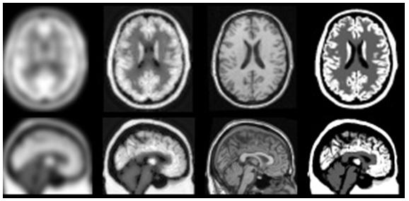
Figure 14. MRI-based partial volume correction for PET images. The first column shows the interpolated PET images. The second column represents the corrected PET. The third column shows the MRI images, and the last column is the ground true of the PET FDG distribution. The axial images are seen at the top, while the coronal slices are seen at the bottom.